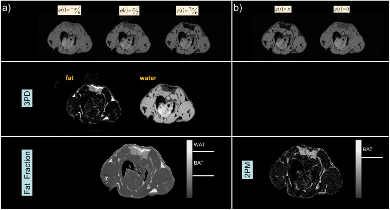
Figure 5. Comparison of the fat-fraction method based on a three-point Dixon approach (3PD, a), and the proposed two-point magnitude based method (2PM, b).
The fat-fraction a) and BAT b) image are normalized from 0 to 1. The grey bars indicate the respective grey values used as threshold for WAT (>0.8, 3PD) and for BAT (>0.4 and <0.8, 3PD; >0.7, 2PM). WAT cannot be identified by the 2PM method, since signal from voxels containing only fat or water will be cancelled out during the involved subtraction.

