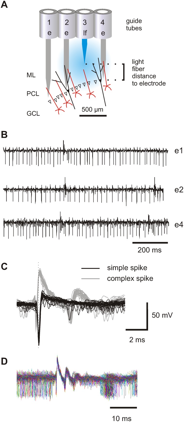
Figure 1. Simultaneous recordings from multiple PCs in the vermis of mice.
(A) Schematic representation of relative positions of three electrodes and a single light guide assembled in a linear array during a recording and stimulation experiment in the cerebellum. The guide-tubes (1 to 4) are part of the multi-electrode system, which allows for independent vertical movement of each fiber. Guide-tubes 1, 2 and 4 each house an electrode while guide-tube 3 contains a light fiber. The electrodes are placed to enable recordings from the Purkinje cell layer, whereas the tip of the light-guide resides in the molecular layer. (ML: molecular layer, PCL: Purkinje cell layer, GCL: granular cell layer). (B) Simultaneous recording from three PCs with electrodes e1, e2 and e4. Complex spikes can be identified by upward deflection of the action potential. (C) Superposition of 30 simple spikes (black) and 30 complex spikes (gray) recorded from electrode e2. (D) Complex spike triggered superposition of raw traces (n = 295) shows that simple spikes do not occur for 15 ms after complex spikes. The simple spike pause indicates that simple and complex spikes are recorded from the same cell.

