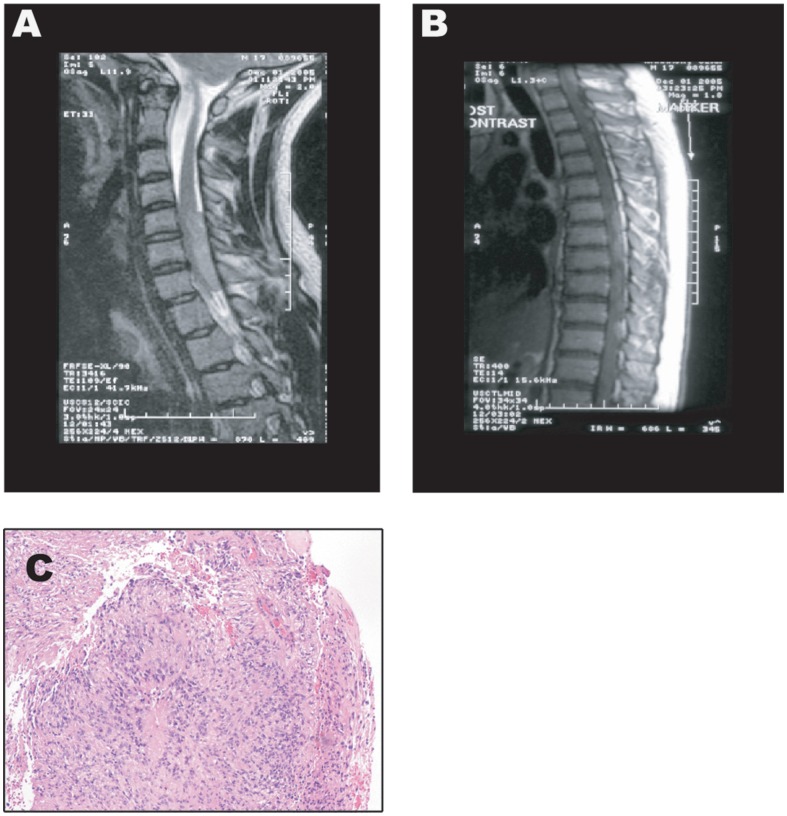
Figure 1. MRI Images and H&E Staining of the Anaplastic Ependymoma Case at Presentation.
A) Magnetic Resonance Imaging (MRI) of the cervical spine showing the presence of an enhancing mass, which extends from mid C5 to inferior C7 (4.5 in length×1.0×2.0 in cephalocaudal and anteroposterior dimension) and causing cord compression. B) MRI of the thoracic spine showing an enhancing lesion at T2–3 (1.5 in length×0.6×0.6 cm in anteroposterior and transverse dimension) with several other smaller nodular masses, best seen on the T2 weighted sequence, which extended throughout the thoracic level to T11. C) Hematoxylin and Eosin staining of a tumor section showing an overall predominant dense cellular component, with primitive nuclear features, mitotic activity, necrosis and vascular proliferation. The presence of well formed, obvious perivascular pseudorosettes (with vasocentric pattern, perivascular nuclear-free zones, and classic thin glial processes radiating to/from the vessel wall) were found supportive of the diagnosis of intradural, extramedullary anaplastic diffuse spinal ependymoma, WHO grade III.

