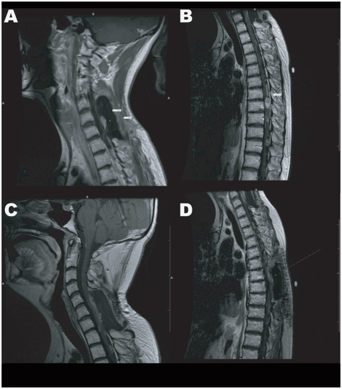
Figure 2. MRI Images of Cervical and Thoracic Spine.
A) 2009 MRI of the cervical spine showing recurrence in the surgical area. B) 2009 MRI of the thoracic spine showing progression of the main lesion measuring 23.9 mm, and the appearance of several other smaller lesions. C and D) 2010 MRI of the cervical and thoracic spine showing tumor regression following a treatment with irinotecan and bevacizumab.

