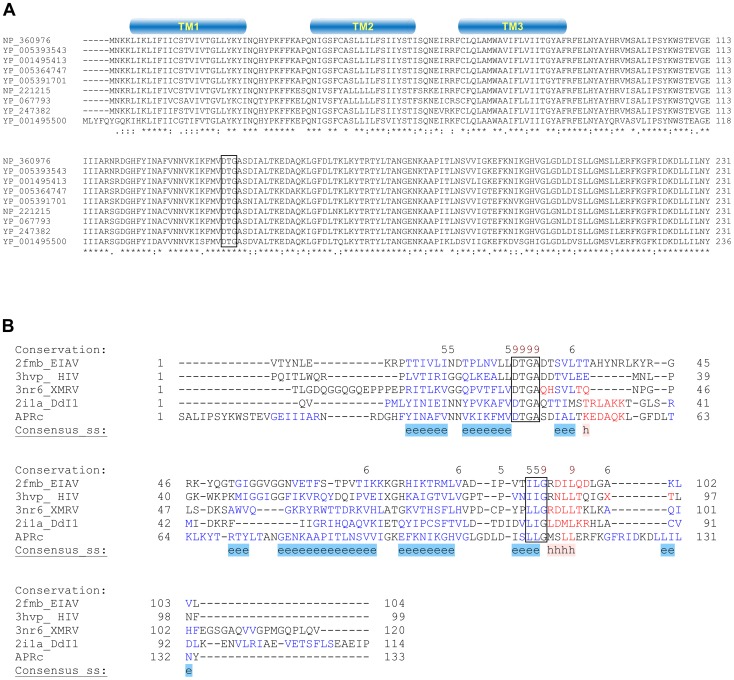
Figure 1. RC1339/APRc gene homologues from Rickettsia spp. display a striking pattern of sequence conservation among each other and retain structural similarity with other members of the retropepsin family.
(A) Multi-alignment of deduced amino acid sequences of the putative retropepsin-like protease from representative species from all rickettsial taxonomic groups (spotted fever group, typhus group, transitional group and ancestral group). Sequences were aligned against RC1339/APRc sequence from R. conorii (NP_360976) using the ClustalW software [69]. Accession numbers and corresponding species are described in Table 1. The predicted α-helical transmembrane domains are represented by cylinders and the box indicates the active site motif (DTG). (B) Structure-based alignment of the soluble catalytic domain of RC1339/APRc with HIV-1 (PDB 3hvp), EIAV (PDB 2fmb) and XMRV (PDB 3nr6) retropepsins and with DdI1 putative protease domain (PDB 2i1a), performed with PROMALS3D [71]. The first line shows conservation indices for positions with a conservation index above 4. Consensus_ss represent consensus predicted secondary structures (alpha-helix: h; beta-strand: e). Sequences are colored according to predicted secondary structures (red: alpha-helix, blue: beta-strand). Red nines highlight the most conserved positions. Active site consensus motif Asp-Thr-Gly and hydrophobic-hydrophobic-Gly sequence are boxed.

