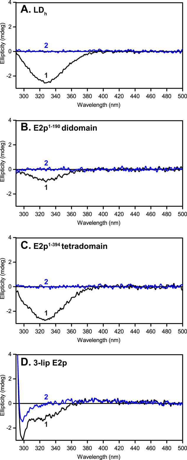
FIGURE 7.

Lipoyl domain has a characteristic negative CD band at 330 nm. Shown are CD spectra of C-terminally truncated E2p proteins and of 3-lipoyl E2p with lipoyl domains in their oxidized state (spectrum 1) and TCEP-reduced form (spectrum 2). A, LDh (0.36 mg/ml). B, E2p(1–190) didomain (0.69 mg/ml). C, E2p(1–394) tetradomain (1.3 mg/ml). D, 3-lip E2p (2.1 mg/ml). The concentration of each protein is ∼30 μm in 50 mm KH2PO4 (pH 7.0) in a total volume of 2.4 ml at 25 °C.
