FIGURE 5.
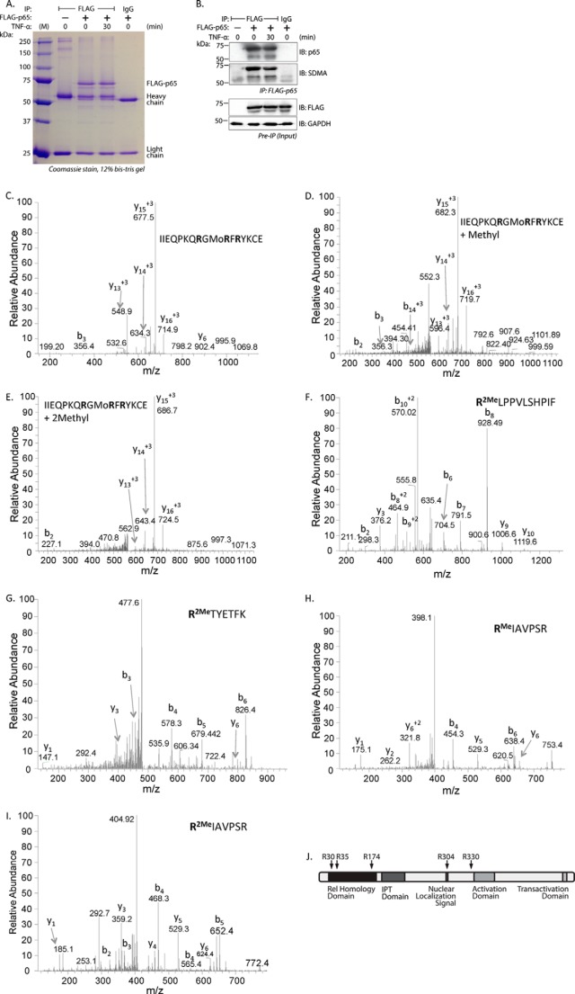
Five arginine residues are dimethylated in EC transfected with wild type p65. A, lysates from wild type p65-transfected EC were immunoprecipitated with anti-FLAG antibody, resolved by SDS-PAGE, and stained with Coomassie Blue. B, top panel, a fraction of the immunoprecipitate in A was probed separately for anti-p65 by immunoblot. The membrane was then stripped and reprobed with anti-SDMA. Bottom panel, preimmunoprecipitated input material was probed with anti-FLAG antibody to show FLAG-p65 expression. The lower molecular mass band in A and B appears to be related to FLAG-p65. C–E, full-length FLAG-p65 bands were analyzed by mass spectrometry for the presence of methylarginine residues. Shown are MS/MS spectra of the unmethylated (C), monomethylated (D), and dimethylated (E) peptide 23IIEQPKQRGMoRFRYKCE39 containing Arg-30 and Arg-35. F, MS/MS spectra of the 174RLPPVLSHPIF184 peptide containing Arg-174. G, MS/MS spectra of the 304RTYETFK310, including Arg-304. H, MS/MS spectra of the 330RIAVPSR336 containing Arg-330. I, MS/MS spectra of the 330RIAVPSR336 peptide. J, the location of methylated arginine residues are shown schematically on human p65. IB, immunoblot; IPT, Ig-like, plexin, transcription factor domain.

