
FIGURE 2.
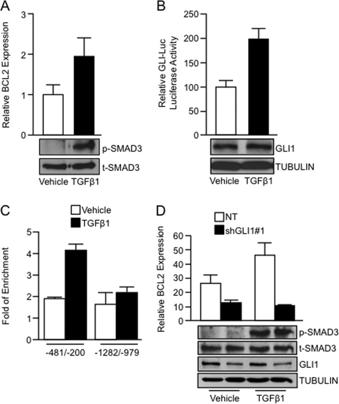
GLI1 is required by TGFβ to regulate BCL2 gene expression. A, real-time PCR showing increased BCL2 mRNA expression in PANC1 cells treated with TGFβ1 ligand. The inset shows increased levels of phospho-SMAD3 (p-SMAD3) in TGFβ1-treated cells, indicative of TGFβ pathway activation. Total SMAD3 was used as loading control. B, PANC1 cells treated with TGFβ1 exhibiting increased GLI-Luc reporter activity compared with vehicle-treated cells. Western blot showed no significant increase in endogenous GLI1 after treatment with TGFβ1 (inset). TUBULIN was used as a housekeeping control. C, ChIP assay was performed using an anti-GLI1 antibody. The data show that upon TGFβ1 stimulation, endogenous GLI1 binding is increased in a region of the BCL2 promoter spanning from −481 to −200 bp upstream the first exon (−481/−200). There was no binding to an upstream sequence in this promoter (−1282/−979). D, real-time PCR showing BCL2 mRNA expression in PANC1 cells transfected with NT vector or GLI1 shRNA targeting construct and treated with vehicle or TGFβ1. The inset shows the levels of the GLI1, phospho-SMAD3 (p-SMAD3), total levels of SMAD3 (t-SMAD3), and TUBULIN. Bar graphs represent average levels in each group ± S.E. (error bars) from three or more replicates.