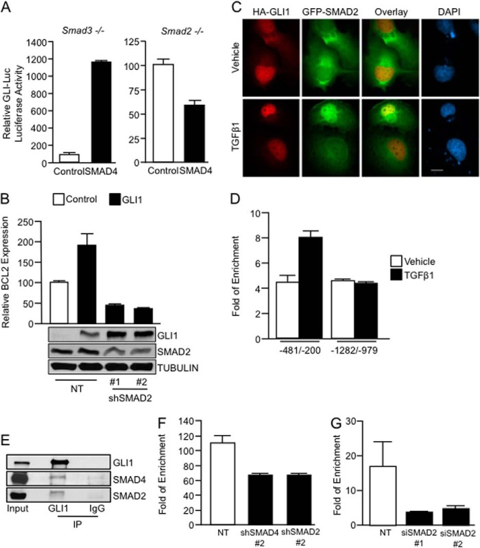
FIGURE 5.
SMAD2 and SMAD4 are required for the recruitment of GLI1 to the BCL2 promoter. A, Smad3−/− and Smad2−/− MEFs were cotransfected with GLI-Luc reporter and either control or SMAD4 expression constructs. Samples were collected for luciferase assay 48 h post-transfection. Basal GLI-Luc activity due to endogenous GLI1 was set at a value of 100 for each control fibroblast line. B, real-time PCR shows BCL2 mRNA expression levels in PANC1 cells transfected with NT or two independent shRNA constructs targeting SMAD2 along with vector control or GLI1 expression constructs. The inset shows levels of expression of GLI1 and SMAD2. TUBULIN was use as housekeeping. Of note is that SMAD2 knockdown does not affect GLI1 expression. C, immunofluorescence in PANC1 cells transfected with HA-GLI1 and GFP-SMAD2 treated with TGFβ1 or control vehicle is shown. D, ChIP assay done in PANC1 cells treated with TGFβ1 shows binding of SMAD2 to the GLI1 binding region of the BCL2 promoter. E, endogenous immunoprecipitation (IP) of GLI1 shows binding of GLI1 with SMAD2 and SMAD4 in RMS13 cells treated with TGFβ1. F, GLI1 ChIP assays in PANC1 cells transfected with the NT, SMAD2, or SMAD4 shRNA and treated with TGFβ1 show that the knockdown of the SMAD2 and SMAD4 factors diminished the binding of GLI1 to the BCL2 promoter. G, similarly, depletion of SMAD2 using two independent siRNAs impairs the binding of SMAD4 to this promoter upon treatment with TGFβ1. Bar graphs represent average levels in each group ± S.E. (error bars) from three or more replicates.

