FIGURE 6.
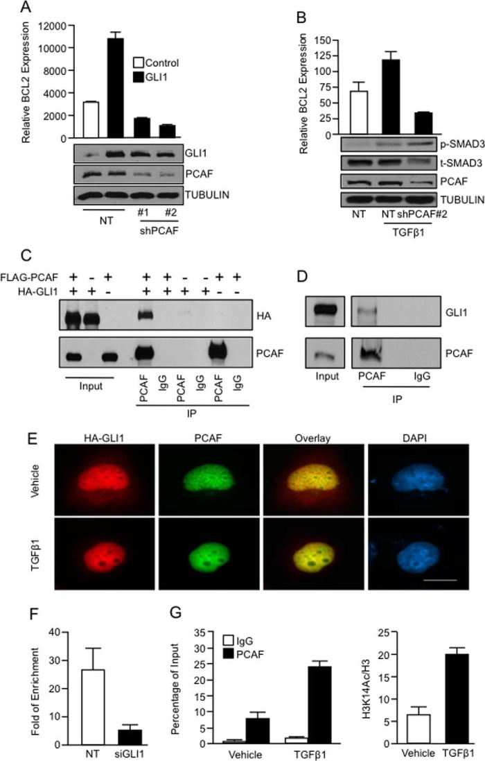
The histone acetyltransferase PCAF is required for activation of gene expression regulated by the TGFβ-GLI1 axis. A, real-time PCR shows mRNA levels of BCL2 in PANC1 cells cotransfected with control or GLI1 expression constructs along with NT or PCAF shRNA vectors. GLI1 expression control by Western blotting showed that the knockdown of PCAF does not affect GLI1 expression. TUBULIN was used as housekeeping control. B, BCL2 mRNA expression is shown in PANC1 cells transfected with NT or PCAF shRNA targeting construct and treated with vehicle or TGFβ1. The inset shows the levels of PCAF knockdown, phospho-SMAD3 (p-SMAD3), and total levels of SMAD3 (t-SMAD3). TUBULIN was use as housekeeping. C, PANC1 cells were cotransfected with FLAG-PCAF and HA-GLI1 and then immunoprecipitated (IP) using an anti-PCAF antibody. Western blot analysis was performed to demonstrate coimmunoprecipitation between GLI1 and PCAF. D, an antibody specific for PCAF was used to immunoprecipitate endogenous GLI1 with PCAF in cells treated with TGFβ1. The immunoprecipitated complex was analyzed by Western blotting. E, PANC1 cells transfected with HA-GLI1 and FLAG-PCAF were treated with TGFβ1 ligand. Immunofluorescence was performed to detect the localization of GLI1 and PCAF using anti-HA and anti-PCAF antibodies, respectively. F, PCAF ChIP assay in PANC1 cells transfected with the NT, or GLI1 siRNA and treated with TGFβ1 showed that GLI1 is required for the recruitment of PCAF to BCL2 promoter. G, ChIP assay performed on PANC1 cells treated with the TGFβ1 ligand or vehicle control shows increase binding of PCAF (left) and an enrichment of histone 3 acetylated lysine 14 (H3K14Ac) in the GLI1 binding region of the BCL2 promoter (right) in cells treated with TGFβ1. Total histone 3 (H3) was used to normalize the levels of H3K14Ac. Bar graphs represent average levels in each group ± S.E. (error bars) from three or more replicates.

