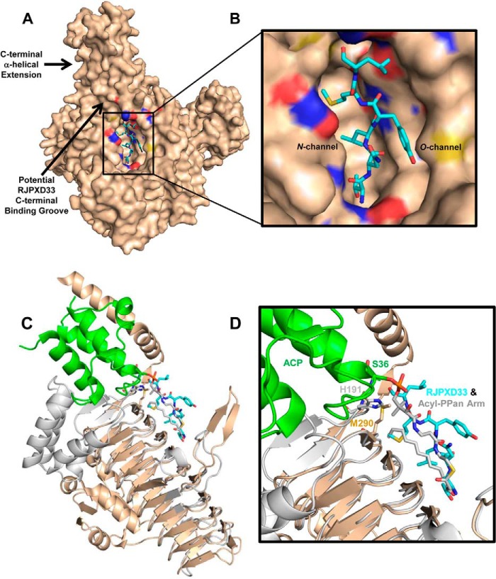
FIGURE 7.
Binding model of the RJPXD33-LpxD complex. A, the EcLpxA-RJPXD33 complex was superimposed onto EcLpxD (PDB code 3EH0; wheat). PyMOL alignment r.m.s.d. = 0.70 over 141 atoms. RJPXD33 (cyan) occupies the fatty acyl binding cleft between two monomers in EcLpxD. The six C-terminal amino acids of RJPXD33 would potentially extend into a binding groove near the C-terminal α-helical extension of EcLpxD. B, close-up view of the putative EcLpxD RJPXD33 binding pocket. C, comparison of RJPXD33-LpxA complex with acyl-ACP-LpxD complex (PDB code 4IHF). Overlay of subunit A of EcLpxA (gray; ribbon) bound RJPXD33 (cyan; sticks) and EcLpxD (wheat; ribbon) bound acyl-ACP (green; ribbon). RJPXD33 occupies the same region as the acyl-PPan prosthetic group (gray; sticks). D, close-up view of the LpxD active site of the EcLpxA-RJPXD33, EcLpxD-acyl-ACP alignment. PyMOL alignment r.m.s.d. = 0.71 over 141 atoms.

