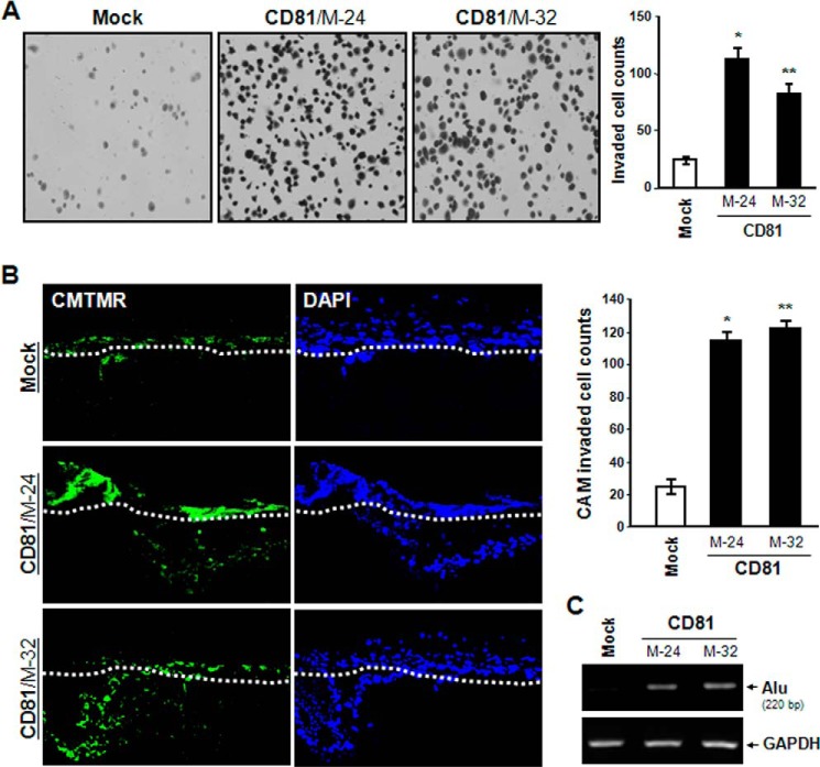
FIGURE 3.
CD81 increases melanoma cell invasiveness. A, cells (2 × 10⁴) were seeded into a Transwell chamber insert equipped with a Matrigel-coated filter. After 12 h of incubation, cells on the lower surface of the filter were stained with Gill's hematoxylin and counted. Results are the mean ± S.D. from three separate experiments with triplicates per experiment (* and **, p < 0.01 versus mock). Error bars represent S.D. B and C, cells were labeled with a fluorescent cell tracker, 5-(and-6)-(((4-chloromethyl)benzoyl)amino)tetramethylrhodamine (CMTMR) (green), and seeded atop the CAM of 11-day-old chicks. After incubating for 3 days, the embryos were frozen and cross-sectioned as described under “Experimental Procedures.” B, CAM sections stained with DAPI (blue) were photographed under a fluorescence microscope. The CAM surface is marked by a dashed line. Cells showing both green and blue fluorescence beneath the CAM surface were counted and expressed as the mean ± S.D. of three sections in each of three embryos (* and **, p < 0.01 versus mock). C, human melanoma cells that penetrated the CAM surface were detected as Alu sequence by PCR on DNA extracted from the lower CAM.

