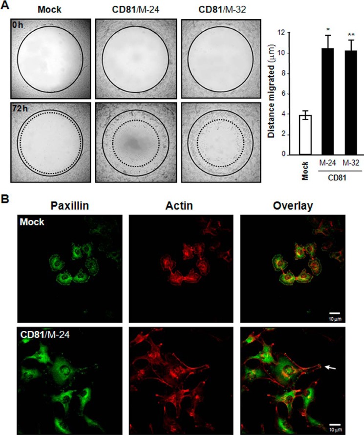
FIGURE 4.
CD81 stimulates melanoma cell migration. A, cell motility was determined using Oris cell migration assay kit as described under “Experimental Procedures.” Shown are representative images taken under a microscope with 40× magnification. Results are the mean ± S.D. from three separate experiments with triplicates per experiment (* and **, p < 0.01 versus mock). Error bars represent S.D. B, cells were permeabilized and incubated with mouse anti-paxillin antibody and Texas Red-labeled phalloidin. Following incubation with Alexa Fluor 488 (green)-conjugated anti-mouse IgG, fluorescence images of cells were taken under a confocal microscope. Shown are the representative images of three separate experiments. An arrow indicates membrane protrusion linked to cell migration.

