FIGURE 3.
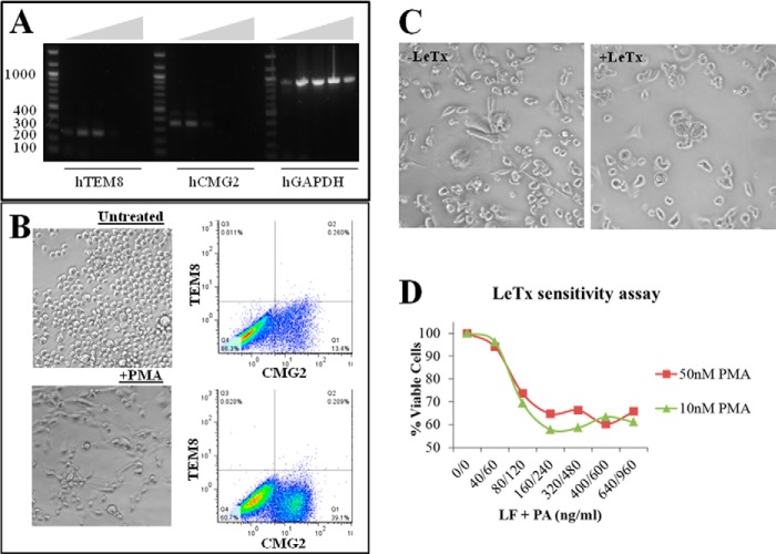
Differentiation of THP-1 cells and LeTx sensitivity. A, total RNA was isolated from THP-1 cells and Superscript III reverse transcriptase was used to synthesize cDNA. Gradient PCR (55–69.1 °C) was used to amplify cDNA fragments corresponding to human TEM8 (256 bp), CMG2 (344 bp), and GAPDH (930 bp). B, comparison of THP-1 (top) and PMA (10 nm)-differentiated THP-1 (bottom) morphology and surface expression of ANTXRs. Brightfield images were taken at ×20,000 magnification using a Nikon Eclipse Ti microscope. C, effects of LeTx in PMA (10 nm)-differentiated (right) compared with untreated cells (−LeTx, left). D, titration of LeTx in THP-1 cells treated with 10 and 50 nm PMA.

