FIGURE 6.

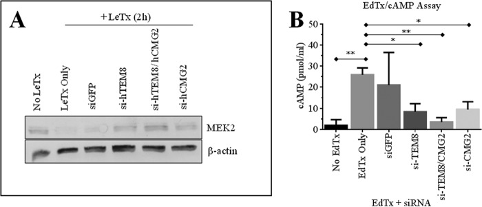
Evaluation of anthrax toxin-mediated MEK2 cleavage and intracellular cAMP in ANTXR-silenced cells. AD293 cells were cultured in 6-well plates and treated with 150 pmol siRNAs for 24 h prior to treatment with anthrax toxins. A, cells were treated with LeTx for 2 h. MEK2 cleavage and β-actin expression were assayed by Western blot. The figure is representative of three separate experiments. B, cells were treated with EdTx (0.25 μg/ml EF + 1 μg/ml PA) for 1 h. Cells were then lysed with 0.1 m HCl, and intracellular cAMP was measured using a commercial ELISA kit. Results (mean ± S.D. (error bars)) from three experiments are shown. One-way ANOVA revealed the groups to be significantly different (p = 0.0057), and Dunnett post hoc comparisons with mock-transfected, EdTx-treated cells (EdTx only) were performed (*, p < 0.05; **, p < 0.01).
