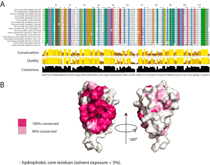
FIGURE 4.
Amino acid residue conservation in SLED domains. A, multiple sequence alignment of the Scml2 SLED domain across the species. Default ClustalW (29) colors are used for each residue. Residues with conservation of 90% or higher are highlighted, and residues that form the hydrophobic core of the domain are annotated with an asterisk. B, SLED surface conservation. Conserved residues (pink) are mapped on the surface of the human Scml2 SLED. Two orientations are shown to illustrate that conserved residues form a continuous patch on the SLED domain surface. The molecule on the right is rotated by 180° relative to the molecule on the left.

