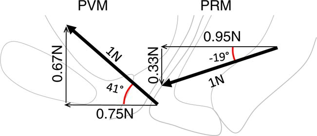
Fig. 3.

Horizontal and vertical components of the PVM and PRM in the standing position. The thick arrows show the average direction of the lines of action of the PVM and PRM muscles relative to the horizontal with a theoretical 1 N force. Thin lines indicate the portion of each force related to a closing and lifting function. (Note: vectors are shown larger than the background anatomy to avoid an overlap in the display)
