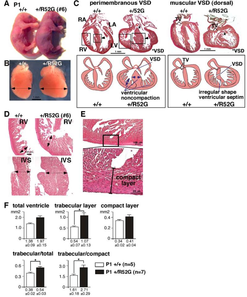
Figure 3. Representative cyanotic cardiac anomaly and noncompaction in P1 Nkx2-5+/R52G mouse in comparison to control wild-type littermate.
Control Nkx2-5+/+ (left) vs. cyanotic Nkx2-5+/R52G mouse #6 (right): (A) whole bodies, (B) dissected hearts, and (C) heart sections with simplified illustrations. (D) Enlarged images of heart sections of right ventricle (RV, top) and interventricular septum (IVS, bottom) obtained from Nkx2-5+/+ (left) vs. Nkx2-5+/R52G mice (right). (E) Enlarged images of tissue sections defining the compact layer morphologically as the appearance of a compact band of uniform tissue, while the endocardial noncompacted trabecular layer consists of trabecular meshwork with deep endomyocardial spaces29. (F) Quantification of area size of total ventricle, trabecular and compact layer. Nkx2-5+/R52G hearts demonstrating profound anomalies were excluded. RA, right atrium; LA, left atrium; RV, right ventricle; LV, left ventricle. *P<0.05.

