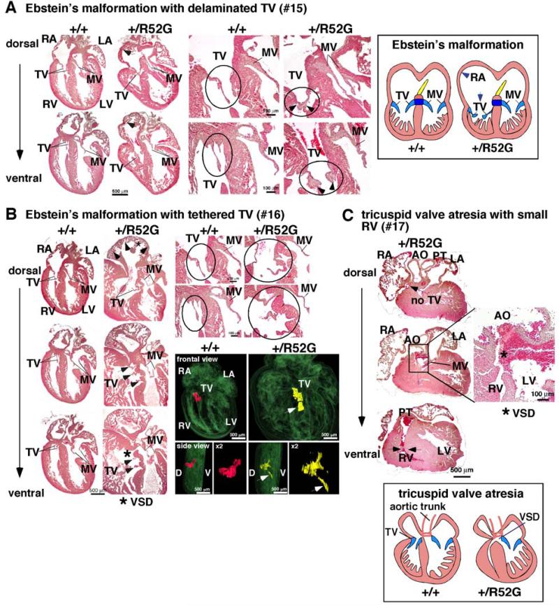
Figure 5. Representative tricuspid valve anomalies in P1 Nkx2-5+/R52G mice.
(A) Ebstein’s malformation in mouse #15. Positions of septal and mural (posterior) leaflets of Nkx2-5+/R52G mice displaced toward apex compared to control mice. Delaminated tricuspid valves are also observed. (B) Ebstein’s malformation with tethered tricuspid valves in mouse #16. 3D reconstructed heart sections including tricuspid valves highlighted in red in control or yellow in Nkx2-5+/R52G mouse. Arrows indicate displaced anterior leaflet of Nkx2-5+/R52G mice toward apex compared to control mice. Of note, septal leaflet could be displaced as well; however it was difficult to distinguish between these two leaflets. (C) Tricuspid valve atresia, muscular VSD, and small right ventricular cavity in mouse #17. See Supplemental Figure S3.

