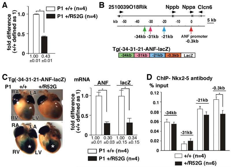
Figure 7. Reduction of ANF and X-gal staining of -34-31-21-ANF-lacZ transgenic mice in Nkx2-5+/R52G hearts relative to those from Nkx2-5+/+.
(A) Difference of ANF mRNA expression normalized to β-actin between Nkx2-5+/+ and Nkx2-5+/R52G hearts. ANF expression in Nkx2-5+/+ hearts defined as 1. Number of animals analyzed is shown. (B) Organization of the genomic locus including ANF (Nppa). Positions of three regulatory elements and the proximal promoter of ANF, and schematic of transgenic lacZ reporter construct are indicated. (C) Representative images of X-gal staining from a total of 4 sets of Nkx2-5+/+ and Nkx2-5+/R52G heterozygous -34-31-21-ANF-lacZ-positive P1 littermates are shown. Decreased lacZ staining in the left ventricle is marked (*). Difference, as determined by TaqMan real-time RT-PCR, between ANF and lacZ mRNA expression normalized to β-actin, with expression in Nkx2-5+/+ defined as 1. Number of animals analyzed is shown. (D) ChIP analysis of Nkx2-5 in percentage of input DNA recovered from 2-3 independent experiments with TaqMan PCR performed in duplicate.

