
Figure 1.
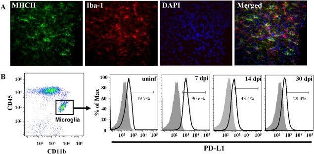
Microglia chronically express MHC Class II and PD‐L1 following MCMV‐induced encephalitis. MCMV‐infected Balb/c mice were perfused (30 dpi) and cryosectioned for immunohistochemistry. (A) Co‐labeling of MHC II (green), as an indicator of activation, and the microglial cell marker, Iba‐1 (red). Nuclear counterstain with DAPI (blue). Tissue section thickness = 25 µm. (B) Mononuclear cells were extracted from uninfected and MCMV‐infected Balb/c mouse brains (7, 14, and 30 dpi). Cells were analyzed for surface expression of PD‐L1 on microglia using flow cytometry. Microglia were identified as CD45intCD11b+ and were specifically gated on for PD‐L1 (black line). Isotype antibody control = filled grey line. Flow plots are representative of three separate experiments.