Figure 2.
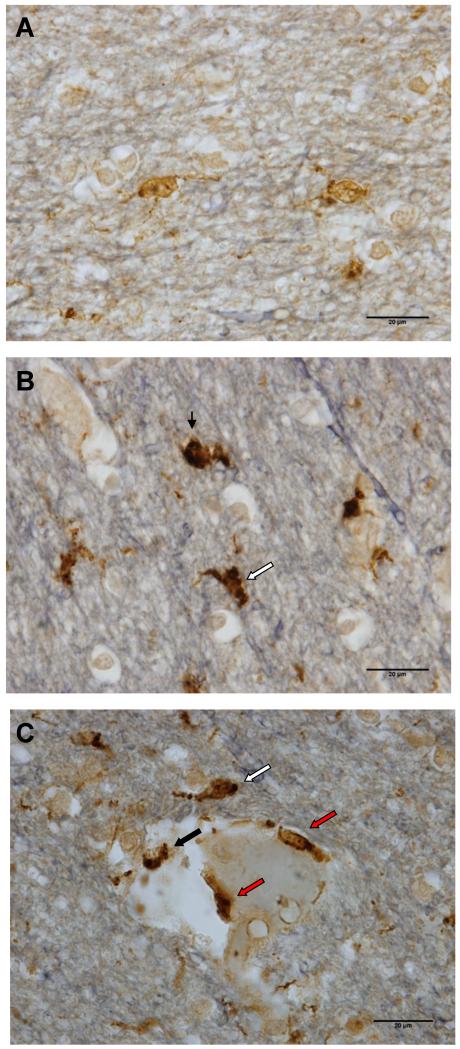
Representative images of microglial phenotypes in human prefrontal white matter, immunostained for Iba-1(brown), CD68 (black) with Verhoeff counterstain (grey-blue). (A) Resting microglia were predominantly Iba-1-immunoreactive with small puncta of immunoreactivity for CD68. (B) An activated microglial cell (white arrow) and a macrophage (black arrow) showed strong immunoreactivity for CD68 (black) obscuring much of the Iba-1 staining (brown). (C) Immunoreactive cells within (red arrows) or in contact with the blood vessel walls (black arrow) were counted as perivascular cells; a cell in close proximity to the blood vessel, but within the parenchyma (white arrow), were counted as activated microglial cells. Similar to activated phagocytes, perivascular cells showed strong CD68 immunoreactivity. Scale bars: 20 μm.

