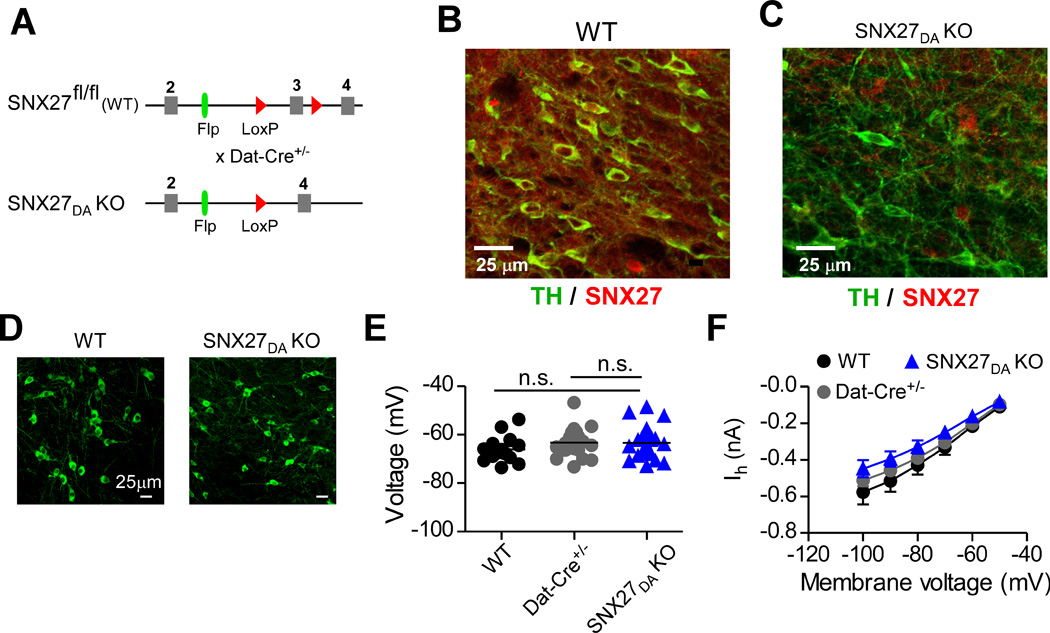
Figure 1. Viability of mice with targeted disruption of SNX27 expression in VTA DA neurons.

(A) Schematic shows conditional deletion of exon 3 in Snx27 occurs in DAT-expressing cells. Floxed animals with SNX27 exon 3 flanked by loxP sites were crossed to DAT-Cre+/− line. SNX27 exon 3 is excised by Cre in DAT-expressing dopamine neurons, referred to as SNX27DA KO mice. (B,C) Dual immunofluorescence for SNX27 (red) and TH (green) in coronal sections from WT and SNX27DA KO. (D) Immunofluorescence for TH in coronal sections from WT and SNX27DA KO. No apparent difference in TH+ density (7.5± 0.99 for WT, n=2 and 6.0 ± 0.26 for KO, n=2 per 150µm2. (E) Resting membrane potentials were indistinguishable in DA neurons from WT, DAT-Cre+/− and SNX27DA KO mice. (F) Ih currents were indistinguishable in DA neurons from WT, Dat-Cre+/− and SNX27DA KO mice.
