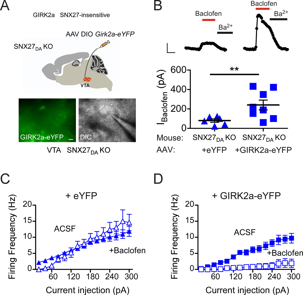
Figure 6. Expression of SNX27-insensitive GIRK2a in VTA DA neurons of SNX27DA KO mice restores GABABR-activated GIRK currents and inhibition.
(A) Schematic shows stereotaxic injection of AAV DIO-GIRK2a-eYFP into VTA of SNX27DA KO mice. Fluorescence and DIC images of eYFP positive cell selected for recording from ex vivo midbrain section of SNX27DA KO mice injected with AAV DIO-Girk2a-eYFP. (scale bar: 20 µm) (B) Top, whole-cell recordings from VTA DA neurons show baclofen-activated GIRK currents (IBaclofen; 300 µM) from WT and SNX27DA KO mice injected with AAV DIO-eYFP or AAV DIO-Girk2a-eYFP, and response to Ba2+ (1 mM). Scale bar is 100 s and 100 pA. Bottom, baclofen-induced currents in GIRK2a-eYFP positive cells are significantly increased from eYFP+ cells (**p<0.05, Student’s t test). Scatter plot of IBaclofen for indicated DA neurons with average current indicated by solid black bar. (C,D) Input-output plots show firing frequency increases as a function of larger current injections in the absence (solid circles, ACSF) and presence of baclofen for DA neurons cells infected with eYFP (n=6) (C) or GIRK2a-eYFP (n=8) (D). Baclofen inhibits firing activity in GIRK2a-eYFP expressing DA neurons (D) but not in eYFP expressing neurons (C) of SNX27DA KO mice (p<0.01 absence versus presence baclofen at 140–300pA, 2-way ANOVA with Bonferroni post hoc test).

