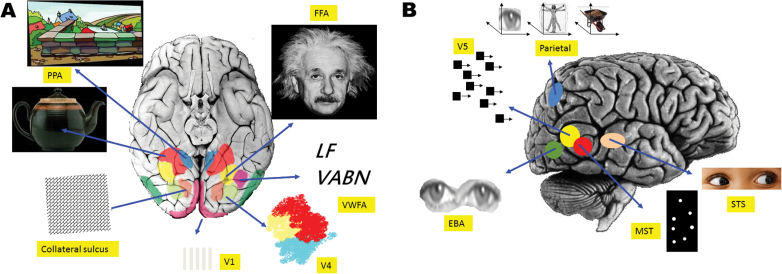
Fig. 3.
Visual cortical specializations. (A) Ventral view of the brain. Selected areas of relative specialization for (from bottom clockwise) contrast, orientation, and luminance (V1); visual texture patterns; objects; landscapes (PPA, parahippocampal place area); familiar faces (FFA, fusiform face area); text and letter strings (VWFA, visual word form area); and colors V4. (B) Lateral view of the brain. Specializations (from left bottom clockwise) for body parts (EBA, extrastriate body part area), motion (V5), parietal eye, body and object reference frames, gaze and social eye signals (STS, superior temporal sulcus), and biological motion (MST).

