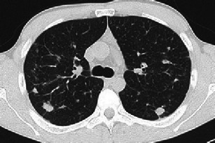
Fig. 5.

A 34-year-old man, smoker, symptomatic for cough and fever. Definitive diagnosis obtained by VATS performed within a month of imaging. A 3-mm collimated CT shows scattered nodules, with asymmetric distribution. The largest nodules measure 15–20 mm; nodular borders are irregular or atypically smooth. Smooth linear interstitial thickening is also evident
