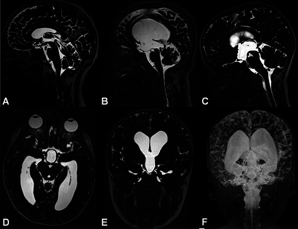
Fig. 1.

Heavily T2W 3D-SPACE images of different cases. A normal midline sagittal image is shown for comparison (A). The rest of the images from two different patients with aqueductal stenosis demonstrate enlargement of the ventricles proximal to the obstruction (B, C), enlargement of the third ventricular recesses (B, C), dilated ventricular horns (D–F) and narrowed cortical sulci (MIP image, E), which are typical findings of obstructive hydrocephalus
