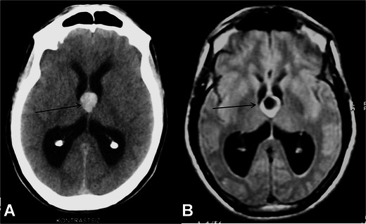
Fig. 3.

A 46-year-old male patient with a colloid cyst. Axial noncontrast-material enhanced CT image demonstrates hyperdense lesions located in the foramen of Monro sized 2 cm in diameter (arrow, A). The lesion is hypointense in FLAIR images, which indicates the proteinous content of the cyst (arrow, B)
