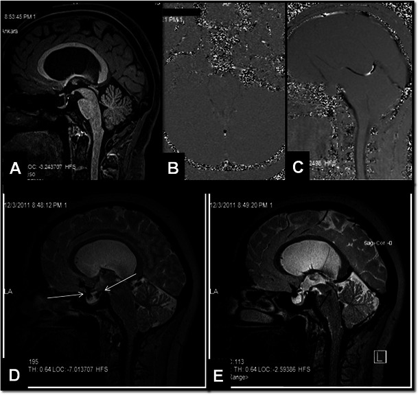
Fig. 4.

A 32-year-old female patient with partial aqueductal stenosis, partial empty sella and hydrocephalus. In the sagittal midline 3D-MPRAGE image no increase in third ventricle size and no aqueductal abnormalities are detected (A). However, axial and sagittal PC-MRI images (VENC value: 6 cm/s) demonstrate lack of CSF flow in the aqueduct (B, C). Sagittal 3D-SPACE image demonstrates passage of CSF from basal-prepontine cisterns into the sellar cavity through the diaphragm sella (white arrows, D). This finding explains why hydrocephalus may be associated with empty sella in most of the patients. In the midline sagittal 3D-SPACE image, a narrowed but patent aqueduct is demonstrated (black arrow, E)
