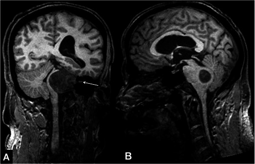
Fig. 5.

Reformatted coronal (A) and sagittal (B) 3D-MPRAGE images of a 50-year-old male patient with a left cerebello-pontine angle mass and hydrocephalus. Coronal image demonstrates the mass extending out of the internal acoustic canal, decompressing and displacing midbrain structures to the right (arrow, A). In sagittal images it is shown that the mass narrows the fourth ventricle and fourth ventricular outlet (B)
