
Fig. 6.
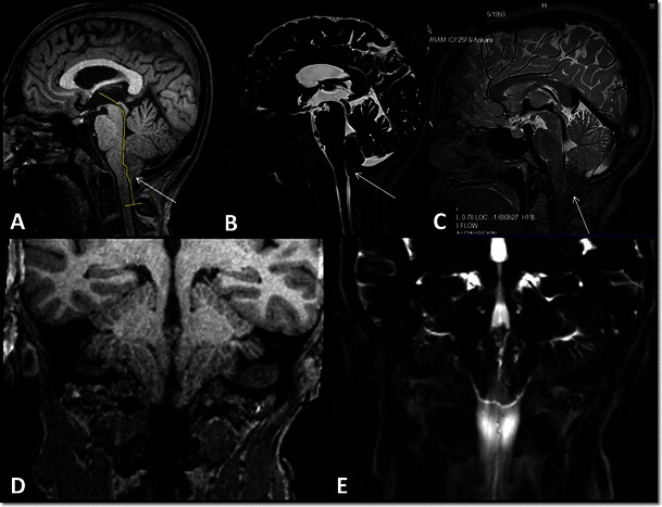
A 19-year=old female patient with Chiari malformation. Sagittal 3D-MPRAGE (A) and heavily T2W 3D-SPACE (B) images demonstrate cerebellar tonsils extending into the foramen magnum (arrows). 3D-SPACE with variant FA mode image shows the narrow foramen of magendi and foramen magnum (arrow, C). Thinned hypointense signal in these foramina is due to decreased CSF flow (arrow, C). On coronal curved reformatted 3D-MPRAGE (D) and heavily T2W 3D-SPACE (E) images, the patency of the foramina and position of the cerebellar tonsils are better evaluated. The coronal curved reformatted images are obtained by drawing a line on sagittal images as in Fig. 6a (yellow line in A)