
Fig. 10.
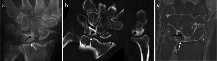
SLAC wrist with further radiological progression (Stage III). Posteroanterior radiograph (a) and coronal and sagittal CT reformat images (b) of the same patient, as well as a coronal STIR MRI image of the wrist (c) in a different patient with SLAC wrist arthropathy. Images demonstrate narrowing of the capitolunate joint (black arrows) in addition to involvement of the radioscaphoid joint. Note preservation of the radiolunate joint (white arrows). There is no significant migration of the capitate at this stage