
Fig. 11.
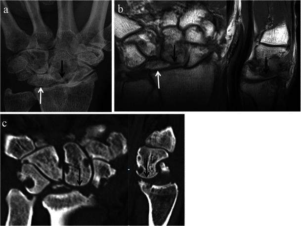
SLAC wrist with late radiological findings. Posteroanterior wrist radiograph (a), and coronal and sagittal T1-weighted MRI images (b) of the wrist in the same patient, and coronal and sagittal CT reformat images (c) of the wrist in a different patient. In addition to extensive osteoarthritic changes, the images demonstrate significant proximal migration of the capitate (black arrows) with ulnar displacement of the lunate (white arrows)