
Fig. 13.
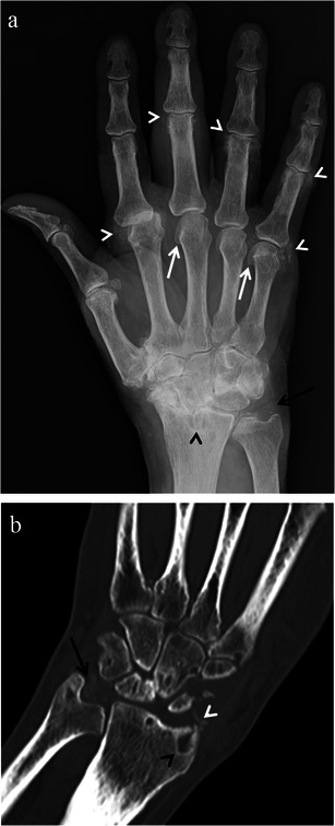
CPPD arthropathy imaging findings. Posteroanterior radiograph of the hand/wrist (a) and coronal reformat CT image (b) of two different patients demonstrating SLAC wrist and classic findings of CPPD arthropathy. These findings include triangular fibrocartilage (black arrows) and peri-articular (white arrowhead) calcifications, prominent subchondral cysts (black arrowheads) and osteoarthritic changes involving the metacarpophalangeal joints including hook-like osteophytes at the metacarpal heads (white arrows). Note is made of significant osteoarthritic changes involving the second metacarpophalangeal joint as well