
Fig. 10.
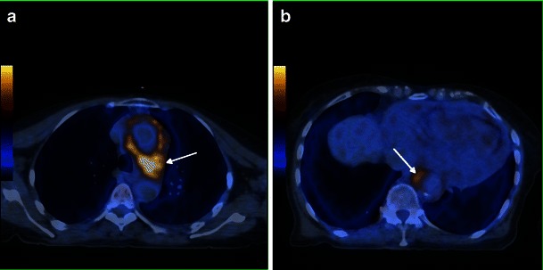
Axial FDG-PET/CT fusion images in a 71-year-old female, in the upper (a) and mid (b) portion of the thorax, showing elevated metabolic activity involving the aortic arch (arrow) and descending aorta (arrow)
Official websites use .gov
A
.gov website belongs to an official
government organization in the United States.
Secure .gov websites use HTTPS
A lock (
) or https:// means you've safely
connected to the .gov website. Share sensitive
information only on official, secure websites.
Axial FDG-PET/CT fusion images in a 71-year-old female, in the upper (a) and mid (b) portion of the thorax, showing elevated metabolic activity involving the aortic arch (arrow) and descending aorta (arrow)