
Fig. 9.
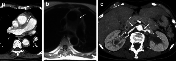
Axial CT-enhanced (a) and axial HASTE MR (b) images of the mediastinum and axial MIP CT (c) image of the upper abdomen in a 73-year-old male. There is periaortic and mediastinal soft tissue infiltration (arrow) and also infiltration of the origin of the renal arteries (arrows) and of the left renal sinus, with mild hydronephrosis (arrow)