Fig. 14.

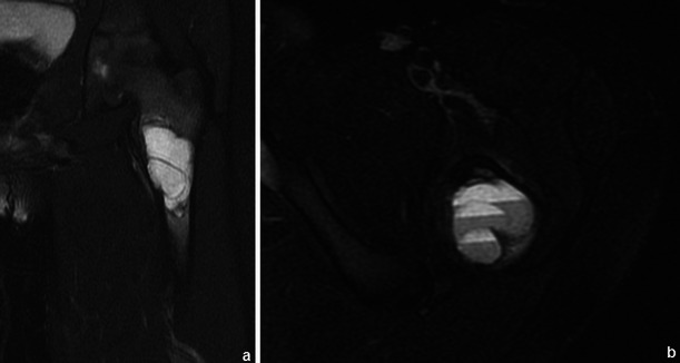
ABC of the left femur: coronal (a) and axial (b) T2FS WI: Typical fluid-fluid levels are seen, with no intervening soft tissue or thick septations. The cortical area is expanded but not disrupted

ABC of the left femur: coronal (a) and axial (b) T2FS WI: Typical fluid-fluid levels are seen, with no intervening soft tissue or thick septations. The cortical area is expanded but not disrupted