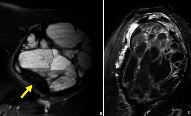
Fig. 15.

Axial T2WI (a) and sagittal post-contrast T1WI (b) show typical features of telangiectatic osteosarcoma: a very large large lesion, with a solid component in the posterior dependent part (arrow in a) and extension beyond the disrupted cortical region (b)
