
Fig. 19.
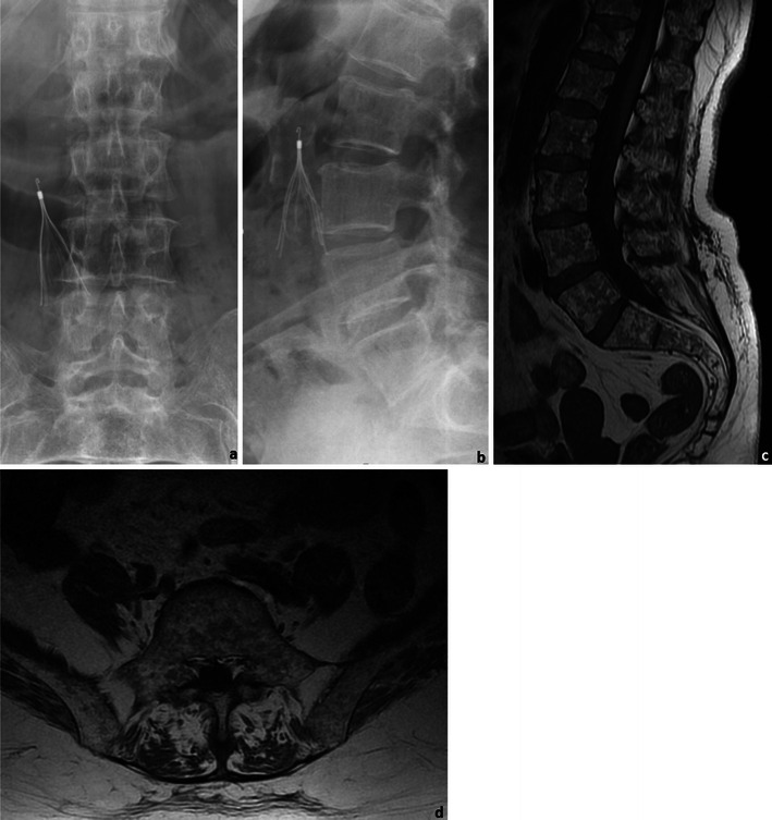
A 76-year-old female patient with MM: Frontal (a) and lateral (b) radiographs of the lumbar spine and sagittal (c) and axial (d) T1WI of the lumbosacral spine. The radiographic study shows diffuse osteopaenia of the lumbar vertebrae, which could only be related to primary osteoporosis given the patients’ gender and age. The MRI study however showed innumerous focal lesions, consistent with the diagnosis of MM. Note: The small cone-shaped device present in the radiographs is an inferior vena cava filter