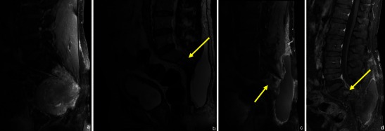
Fig. 26.

MRI applied in the diagnosis and follow-up of a patient with chordoma: this patient had a voluminous lesion destroying the distal part of the sacrum (a). He had radiotherapy and surgery and was re-evaluated 4 months later: MRI showed a posterior subcutaneous fluid collection and a localised area with high T2WI signal (arrow in b) and contrast enhancement (arrow in c) in the inferior spine, which could be due to either residual tumoral tissue or radiotherapy. Posterior follow-up however showed a localised round area of persistant enhancement, which proved to be local recurrent/residual tumour (arrow in d)
