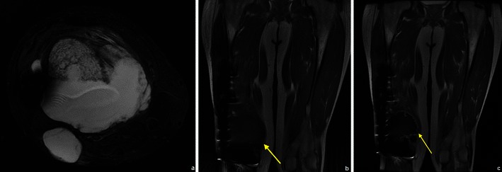
Fig. 27.

This patient had resection of the distal femur due to a chondrosarcoma. MRI showed a suspicious area of high signal near the distal prosthesis (a and arrow in b), which showed peripheral enhancing lobulations (arrow in c): a local recurrence of chondrosarcoma
