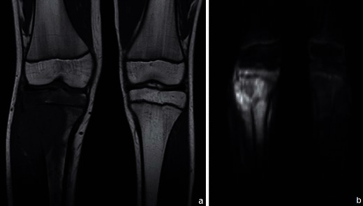
Fig. 3.

Osteosarcoma of the tibia: coronal T1WI (a) shows a large hypointense lesion in the proximal tibia, which breaks through the bony cortex and invades the adjacent soft tissue. Corresponding DWI (b = 700 s/mm2) (b) reveals restricted proton diffusion, consistent with malignancy. Tumoral necrosis (in the centre of the tumour) displays a less hyperintense signal. (Images published by Markus U., Reichardt W. and Kontny U. in MRI: New Developments in Bone Tumor Imaging. Magnetom Flash 2/2011© used with permission of Prof. U. Markus)
