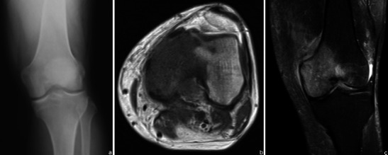
Fig. 4.

Patient with chronic left knee pain. Radiographs were obtained (a normal frontal radiograph). Subsequent MRI study showed abnormal signal intensity in the bone: a low T1WI signal (b) and T2FS WI hyperintense lesion (c) related to a bone lymphoma
