Fig. 7.

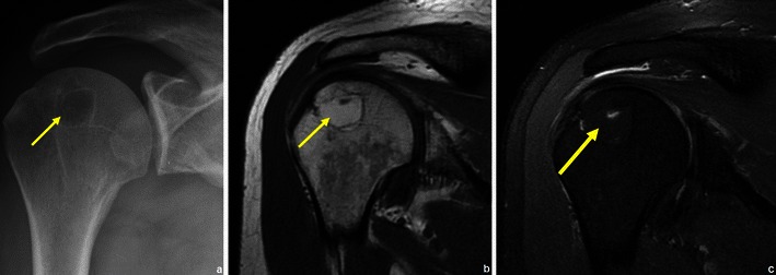
Patient with a humeral lipoma (arrows in a, b and c): frontal radiograph shows a lytic lesion in the head of the humerus. This lesion showed signal intensity similar to that of fat on T1WI (b). The signal was suppressed in T2FS WI (c)

Patient with a humeral lipoma (arrows in a, b and c): frontal radiograph shows a lytic lesion in the head of the humerus. This lesion showed signal intensity similar to that of fat on T1WI (b). The signal was suppressed in T2FS WI (c)