Figure 5.
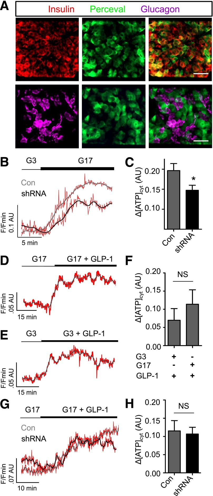
ADCY5 alters β-cell energetics. A: Expression of the ATP/ADP probe Perceval is predominantly restricted to β-cells, as shown using immunohistochemistry with antibodies against insulin and glucagon (scale bar, 25 µm [top panels] and 20 µm [bottom panels]). B: Glucose (17 mmol/L) (G17) still induces increases in ATP-to-ADP ratio after ADCY5 silencing (representative traces shown; gray/black, smoothed; red, raw). C: Bar graph showing a significant effect of shRNA treatment on the amplitude of cytosolic (cyt) ATP/ADP rises in response to 17 mmol/L glucose (*P < 0.05 vs. Con; Mann-Whitney U test; n = 9–10 islets from three donors). D: GLP-1 increases ATP/ADP in the presence of permissive (17 mmol/L) glucose concentrations (representative traces shown; gray/black, smoothed; red, raw). E: As for D, but in the presence of nonpermissive (3 mmol/L) glucose concentrations. F: Summary statistics demonstrate similar effects of GLP-1 on ATP/ADP in islets exposed to 3 or 17 mmol/L glucose (NS, nonsignificant vs. Con; Mann-Whitney U test; n = 10–13 recordings from five donors). G: ATP/ADP responses to GLP-1 are similar in Con and ADCY5 shRNA-treated islets (representative traces shown; gray/black, smoothed; red, raw). H: Summary statistics demonstrate no significant effect of gene silencing on GLP-1–induced ATP/ADP rises (NS, nonsignificant vs. Con; Mann-Whitney U test; n = 6 recordings from two donors). Values represent mean ± SEM.

