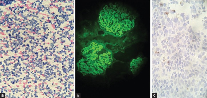
Figure 4.

Digital images captured from various types of specimens. (a) Cytology specimen showing lymphoma captured at high power using an iPhone and the Skylight adapter with digital zoom (Papanicolaou stain, ×400). (b) Fluorescence in situ hybridization (FISH) for IgG in a renal biopsy captured using an iPhone and the Magnifi adapter (IgG FITC, ×200). (c) In situ hybridization for HPV at 400 × captured with a Motorola Droid using Snapzoom adapter (HPV in situ hybridization, ×400)
