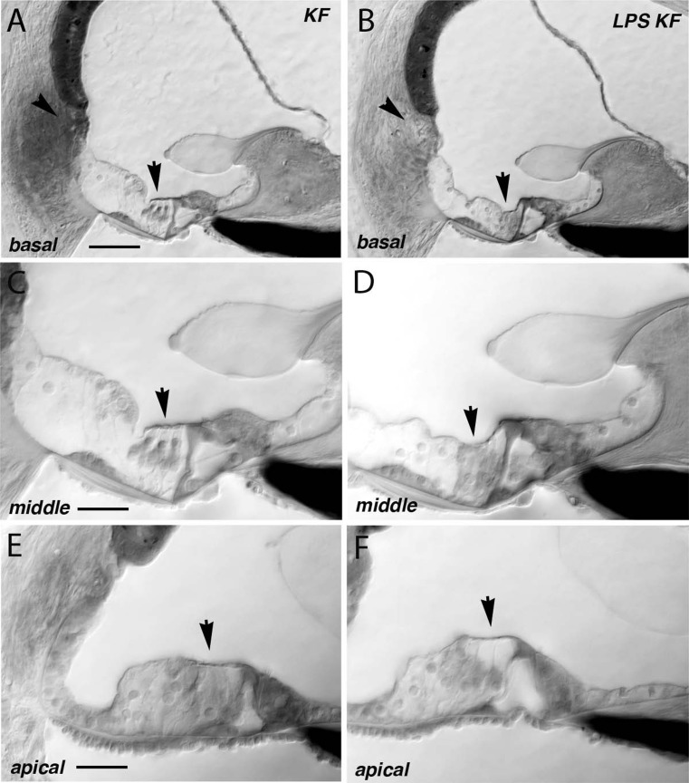
FIG. 7.
Representative sections of the mouse cochlea after kanamycin-furosemide and LPS-kanamycin-furosemide. Plastic-embedded mouse cochleas were sectioned and analyzed for histologic changes after treatment. Representative sections of the mouse basal turn (A), middle turn (C), and apical turn (E) after kanamycin-furosemide. In this specimen, the basal turn showed preserved hair cells while the apical hair cells were absent. The organ of Corti was otherwise preserved, and the inner hair cells were present. B, D, F representative sections after LPS-kanamycin-furosemide treatment. All turns showed complete loss of outer hair cells. In other specimens in this series, we observed sporadic retention of outer hair cells mostly in the basal half. Scale bar in A 50 μm (applies to A, B), C 25 μm (applies to C, D, E, and F). Arrows indicate area of outer hair cells.

