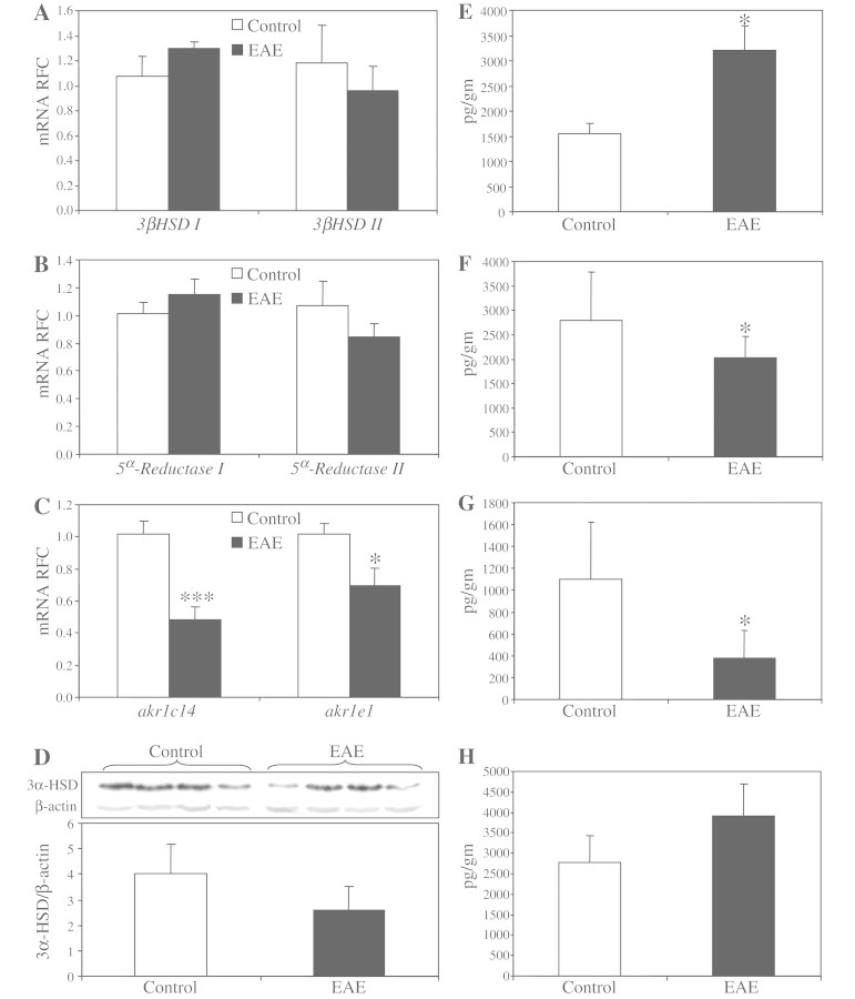
Figure 6.
Neurosteroid biosynthetic machinery and neurosteroid levels are dysregulated in experimental autoimmune encephalomyelitis. Similar to multiple sclerosis brains, real-time reverse transcription–PCR did not show any differences for the transcript levels of 3β-hydroxysteroid dehydrogenase in the hindbrains of experimental autoimmune encephalomyelitis mice (A). Likewise, the transcript levels of 5α-reductase 1 were not changed in multiple sclerosis brains, while 5α-reductase 2 showed a non-significant reduction in experimental autoimmune encephalomyelitis hindbrains (B). In contrast, the two most abundant isoforms of akr1c/e, i.e. akr1e1 and akr1c14, were significantly suppressed in experimental autoimmune encephalomyelitis hindbrains (C). Western blot analysis of EAE hindbrains displayed reduced 3α-hydroxysteroid dehydrogenase (3α-HSD) immunoreactivity in protein lysates derived from EAE tissues (D). Gas chromatography–mass spectrometry of neurosteroid levels showed enhanced levels of pregnenolone (E) in EAE-derived CNS tissues. However, the levels of dehydroepiandrosterone (F) and allopregnanolone (G) were significantly suppressed in EAE hindbrains, whereas the levels of the 3β,5α-tetrahydroprogesterone (THP) isoform were unchanged (H). Transcript levels are represented as relative fold change mean ± SEM. All tissues were extracted from the animals at the peak of the disease (Days 16–17 post immunization). Neurosteroid levels are shown as mean ± SEM (EAE, n = 9 and controls, n = 9; Student's t-test; *P < 0.05, ***P < 0.005; mRNA = messenger RNA; RFC = reduced folate carrier).

