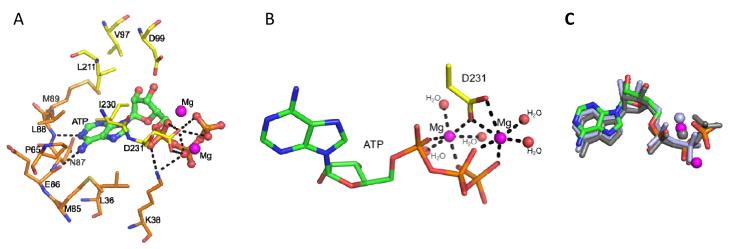
Fig. 3. Nucleotide binding by EhIP6KA.
A, ATP is depicted as a stick and ball model. Two Mg atoms are depicted as magenta spheres. Polar contacts are shown with dashed lines. Amino acids are shown as stick. B, Metal coordination. Two Mg atoms are depicted as magenta spheres. Water molecules are depicted as red spheres. The structure of ATP and Asp231are shown as stick models. Polar contacts to coordinate with Mg atoms are shown with dashed lines. C, The orientation of the EhIP6KA-bound nucleotide (green for carbon, red for oxygen, blue for nitrogen and orange for phosphorus atoms) and Mg atoms (magenta spheres) are superimposed upon that for HsIP3KA (grey stick represent AMPPNP (ATP analog); grey spheres represent Mg), and ScIPMK (light blue stick and spheres represent ADP and Mg).

