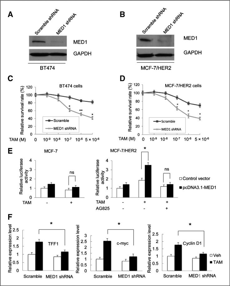
Figure 3.
Knockdown of MED1 sensitizes HER2 overexpression cells to tamoxifen treatment. A and B, Western blot analyses of MED1 levels in BT474 and MCF-7/HER2 cells after control scramble or MED1 shRNA treatments. C and D, control or MED1 shRNA knockdown BT474 cells (C) or MCF-7/HER2 cells (D) were treated with vehicle (Veh) or indicated amount of TAM for 7 days. Cells were then harvested and assessed for cell proliferation by MTT assays. E, MCF-7 and MCF-7/HER2 cells were transfected with vector control pcDNA3.1 or pcDNA3.1-MED1, along with plasmids expressing ERE-TK-LUC reporter and PRL-TK (internal control), followed by vehicle (ethanol) or 1 μmol/L TAM treatment for 24 hours. The relative luciferase values are expressed as mean ±SE. F, real-time RT-PCR was conducted to determine TFF1, Myc, and cyclin D1 mRNA levels in control scramble or MED1 shRNA knockdown MCF-7/ HER2 cells after normalization to that of GAPDH. (* , P < 0.05; ** , P < 0.01).

